![]()
Welcome to MCLinc’s April 2015 Newsletter
Spring has finally arrived in East Tennessee. The beautiful weather makes everyone feel better and gives us cause for celebration. Here in the western part of Oak Ridge there is much to celebrate with the advent of the Manhattan Project National Historical Park. Sites in Oak Ridge, Tennessee, Los Alamos, New Mexico and Hanford, Washington have been designated as a new three-site National Park. Congratulations to those who have worked diligently to bring this memorial to the Manhattan Project to a reality.
The West has another new landmark – the world headquarters of CVMR Corporation. MCLinc is pleased to welcome our new neighbor and looks forward to working with them. It took a Community to bring this important economic development to this area. Many thanks to all who played a role in this recruitment.
In this issue of our newsletter, we feature MCLinc’s capabilities in electron microscopy. A couple of events brought this capability to the forefront – the installation of MCLinc’s newest electron microscope and MCLinc’s recognition by ORNL as an Innovator. We invite you to read more about how these two events are related.
MCLinc continues support of local charitable causes – CASA, the Literary Luncheon, Boys & Girls Club capital campaign, Roane County STEM robotics, ADFAC’s and ORUD’s “Have a Heart, Heat a Home” and more. We also find time to appreciate our friends and customers. Join us when you can.
Thank you for your support.
Barry
MCLinc Acquires New
Transmission Electron Microscope (TEM)
Electron microscopy is a material analysis technique that utilizes a focused beam of electrons to evaluate specimens of interest. In general, two types of electron microscopes exist: scanning electron microscopes (SEM) and transmission electron microscopes (TEM). SEMs generate images by collecting secondary electrons generated through interactions of the electron beam with the specimen’s surface, or back scattered electrons reflected back by the charged subatomic particles that comprise the specimen. TEMs, like MCLinc’s new JEOL 2010 TEM shown below, instead generate images by collecting electrons that have passed through a thin specimen.

Figure 1. MCLinc’s JEOL 2010 Transmission Electron Microscope (TEM).
Both types of electron microscopes are capable of attaining nanoscale or better resolution of microscopic structures, allowing for analysis of topography and morphology. In general, TEMs offer better resolution at higher magnifications over SEMs, and they have found use in numerous fields, ranging from material research, geology, and metallurgy to applications in the medical field, biology, nanotechnology, and forensic analysis. Transmission electron microscopes are regularly used to study the structures of crystalline materials and the morphology of metals and to assess the production and manufacturing of semiconductors and materials such as carbon fiber, where their abilities can be used to assess micro-scale flaws and defects in parts. Interactions between the electron beam and the sample also generate x-rays of characteristic wavelengths dependent upon the elements present within the sample, allowing for compositional analysis of an unknown specimen when used in tandem with tools such as energy dispersive x-ray spectroscopy (EDS). Using the imaging capabilities of the TEM and the elemental data gathered by EDS it is possible to identify asbestos fibers within a sample. Below, images of two types of asbestos, chrysotile and tremolite, are shown. EDS is the key factor to distinguishing between the two materials because they appear very similar in structure, but possess different x-ray spectra.

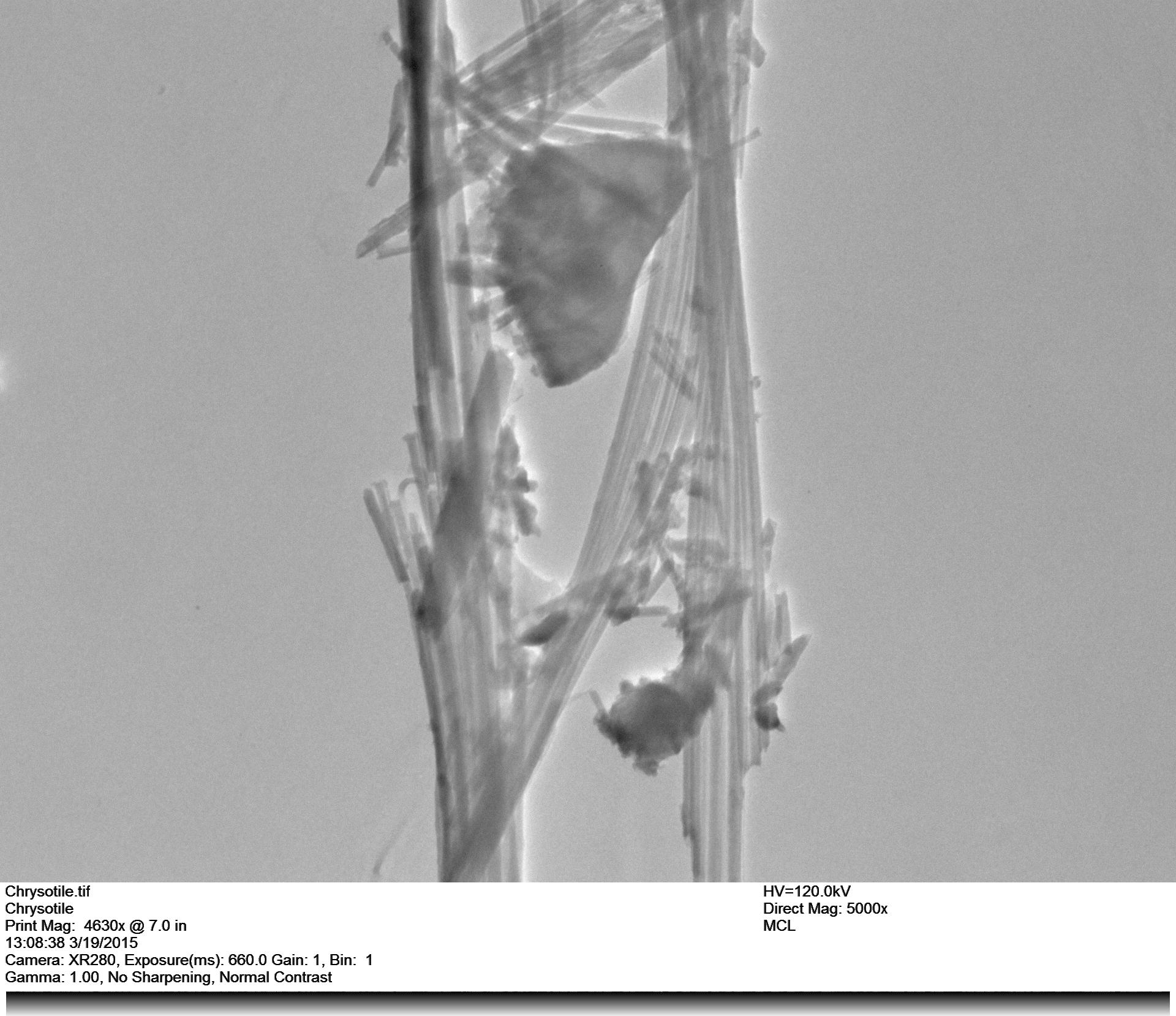
Figure 2. TEM Image of Chrysotile Asbestos Figure 3. TEM image of Tremolite Asbestos
Additionally, diffractions patterns may be obtained by adjusting the lenses of a TEM. These patterns can be used to gather information on the crystalline nature of the specimen along with information of the specimen’s lattice orientation, with respect to the electron beam, and identify the space group symmetry of the material being analyzed. For example, the regular pattern of spots shown in the image below identify the material, which is amosite asbestos, as crystalline in nature. All of these features and more make the transmission electron microscope a versatile tool for research and analysis.
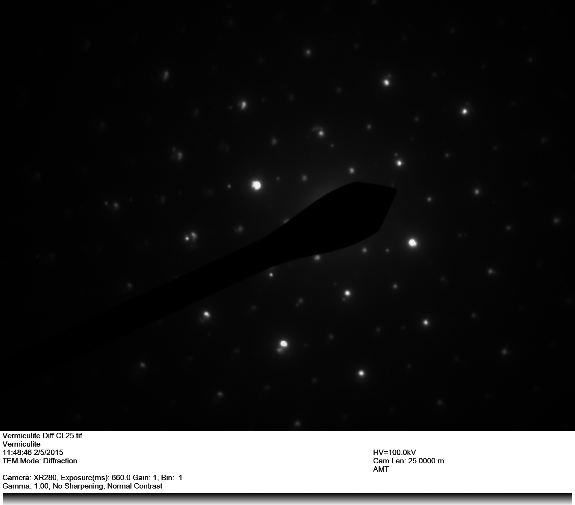
Figure 4. Electron diffraction pattern for Amosite (Grunerite) Asbestos
Imaging Carbon Fibers at MCLinc
Carbon Fiber is a strong, lightweight material used in the manufacture of many products. The characteristic of being lightweight results from the low atomic weight of carbon, and its strength is derived from the arrangement of long, graphitic crystals aligned parallel to the length of the fiber. Carbon fiber is usually manufactured from a synthetic fiber precursor such as rayon or polyacrylonitrile (PAN). The precursor fiber is heated to 300ºC in air, thus oxidizing the material. The oxidized fibers are then heated to 2000ºC in an inert (argon, or nitrogen) atmosphere to induce graphitization of the material. The result is a fiber consisting of graphene sheets aligned parallel to the long dimension of the fiber. The chemical bonds connecting individual carbon atoms in the graphene sheet are exceedingly strong. The strong carbon-carbon bonds contribute to the high tensile strength of carbon fiber.
Carbon fiber manufacturers routinely measure tensile strength of their products using physical testing methods. However, manufacturers also want to know the causes of variations in tensile strength and need methods which can detect and document defects in carbon fiber that can reduce fiber strength. Direct observation of carbon fiber (and precursor) defects can be made using the microscopy facilities at Materials and Chemistry Laboratory, Inc. (MCLinc). Being transparent, the internal features of precursor PAN fibers can be imaged using polarized light microscopy (PLM), which is an optical microscopy method. Because carbon fiber is opaque, its internal features can’t be imaged using PLM. The internal features of carbon fiber can be imaged using Transmission Electron Microscopy (TEM). Carbon fibers typically have a diameter of 5 to 10 microns. For most materials, this would be too thick to transmit electrons through the material. However, since carbon has a low atomic number, carbon has a low electron density. Because of this, a high energy electron beam can penetrate a carbon fiber so that internal features can be directly observed. Surface features of both precursor material and carbon fiber can be imaged using scanning electron microscopy (SEM).
The MCLinc microscopy department is equipped with a full line of optical microscopes, two SEM instruments (JEOL 840 and Hitachi 4500), and two TEM instruments (JEOL 2000EX and JOEL 2010). MCLinc’s newest instrument, the JEOL 2010 TEM, is a high power (200Kv) high resolution instrument that provides high resolution images to 1,000,000X magnification. With this new TEM, MCLinc can now offer a full range of microscopy services to the carbon fiber industry. The following example images, a sort of annotated guide to carbon fiber microscopy, illustrate some of what MCLinc can offer to the carbon fiber industry.

Author of the Imaging Carbon Fibers article is MCLinc employee, Mark R. Colberg, Ph.D. Dr. Colberg (left) is the Technical Manager for Microscopy and Project Manager at MCLinc. He has a B.S. degree in Geology with a minor in Chemistry from Auburn University; a M.S. degree in Geology from Auburn University and a PhD in Geology from the University of Georgia.
Author of the Transmission Electron Microscope (TEM) article is MCLinc employee, Blake Dunaway (right). Blake obtained a B.S. in Material Science and Engineering from the University of Tennessee, Knoxville. He graduated from the Georgia Institute of Technology with his M.S. degree in Bioengineering/Materials Science and Engineering.
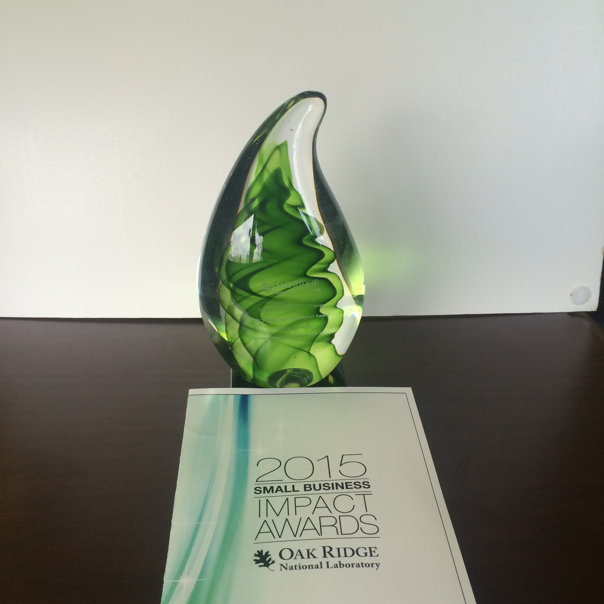
ORNL Small Business Impact Award.
ORNL team who nominated MCLinc for the Innovator award. Back row left to right: Jim Schubert, Derek Miller, Karoly Magda.
MCLinc is very honored to have received the 2015 Oak Ridge National Laboratory Small Business Impact Award in the “Innovator” category. This award honors a small business that has developed an outstanding idea, process, project management mechanism or technology that proved crucial to the success of the ORNL client’s project.
MCLinc won the Innovator Award for developing a process to analyze water filters from a recirculating water system at ORNL. For several years, MCLinc has monitored filters at intervals to characterize materials trapped by the filters to reveal the origin of the material(s). Early in the project, MCLinc suggested using Scanning Electron Microscopy – Energy Dispersive Spectroscopy (SEM-EDS) to examine the water filters because this technique reveals shape and size of particles found in the filters along with the elemental composition of individual particles. Particles were subsequently analyzed by X-ray Diffraction Spectroscopy (XRD) which gives insight into their origin from erosion of a surface versus corrosion in the system. Results of the monitoring have been used to predict impending pump failure prior to their actually shutting down the system.
CASA BBQ and
Bluegrass Bash

MCLinc congratulates CASA of the Tennessee Heartland on the success of the 12th annual CASA BBQ and Bluegrass Bash. The event was held on January 27, at a new location, 205 Main Event Center, located at 205 Main Street in Clinton, Tennessee.
Wonderful music was provided by the Ridge City Ramblers. Those who braved the ice and snow were rewarded with an evening was full of delicious BBQ, and silent and live auctions. MCLinc is proud to be a corporate sponsor of CASA.
If you would like to know more about CASA, or how you can get involved, please follow this link for more information.
MCLinc Supports Boys & Girls Club
The Boys and Girls Club of Clinch Valley operates two campuses: the Lawrence A. Hahn Club in Oak Ridge and the Roane County Club located at Harriman Middle School. The organization’s mission is to enable young people, especially those who need it most, to reach their full potential as productive, caring, responsible citizens. The Oak Ridge Unit and organization’s operations are based out of the Lawrence A. Hahn building in Oak Ridge, named after the Club’s founder and Executive Director Emeritus, Lawrence Hahn, who remains an active volunteer.
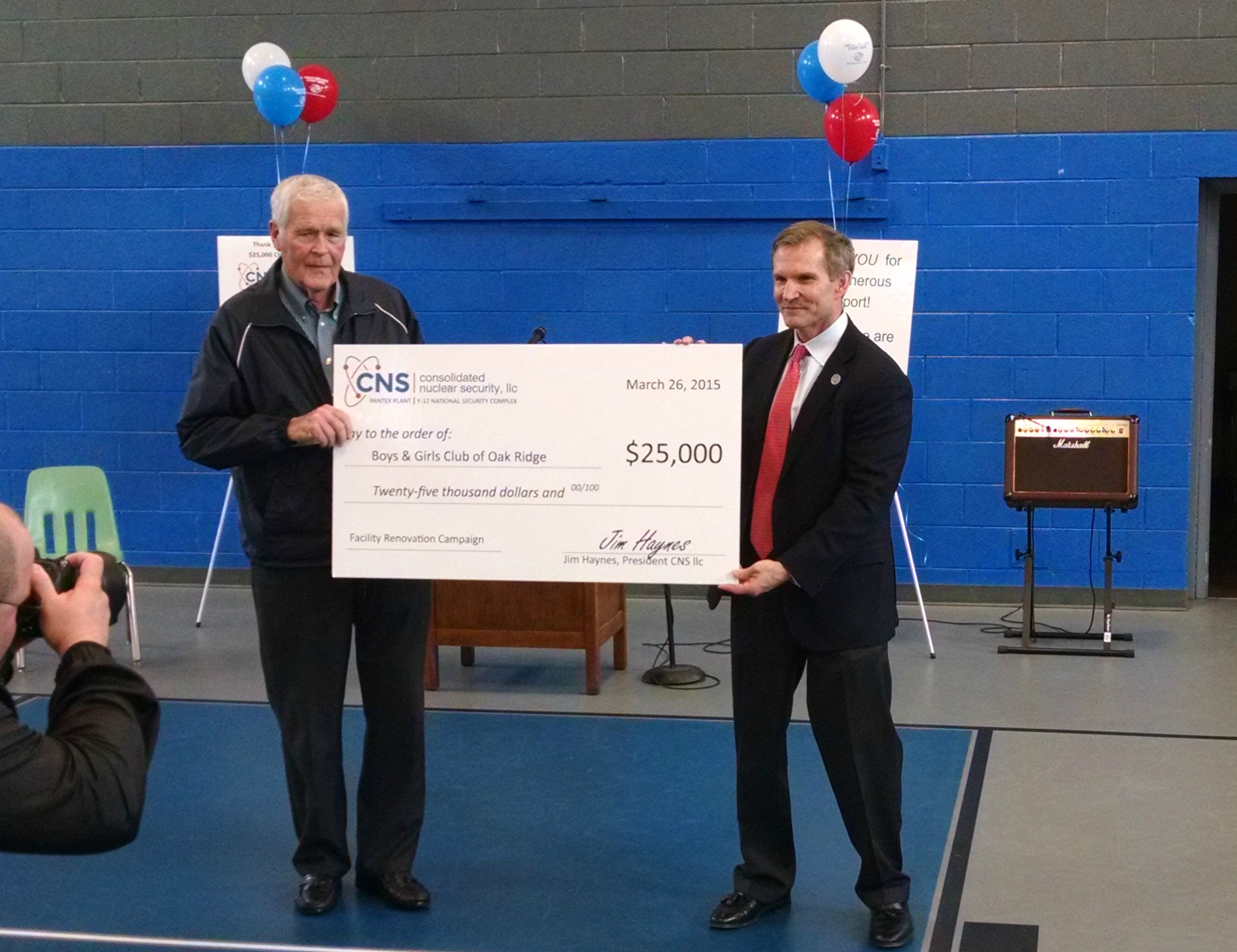
Pictured above, Jim Haynes of CNS presenting a check to Lawrence Hahn of the Boys and Girls club for much needed renovations to their current building. CNS did a $25,000 matching program of which MCLinc donated $1,000.
The Boys and Girls club invites you to tour the facility to “see the need” and consider how you can become part of the great future of the Boys and Girls Club. For more information, or to download a pledge card, please visit bgcor.org or call Jennifer Pettyjon at (865) 244-6073 to learn how you can get involved.
MCLinc Customer Appreciation Pancake Breakfast

MCLinc’s Thomas Dyke and his Heart Shaped Pancakes.
Thanks to all who attended the annual MCLinc Customer Appreciation Pancake Breakfast on February 12th.
The morning was filled with wonderful food and company as the MCLinc crew prepared a delicious breakfast of pancakes, sausage, eggs, and bacon for their guests. The event provides an opportunity for the MCLinc staff to meet our customers and say “Thank You” for their business with MCLinc. In addition, the casual atmosphere at the Heritage Center provided an opportunity for attendees to visit with each other.
MCLinc again invited Oak Ridge Utility District (ORUD) to sell Valentines gifts at the event to support the “Have a Heart, Heat a Home” campaign. All proceeds make it possible for needy families to keep their heat on during the winter months. MCLinc appreciates ORUD’s work on this worthwhile mission. Many thanks to all who made contributions.
MCLinc will be hosting a Customer Appreciation Ice Cream Social, featuring homemade waffle cones, in July. Watch your e-mails for more information. We hope to see you there!
Supporting Robotics

MCLinc helped Roane County in raising funds for their STEM Robotics team. Roane County High School first established a robotics team in 2013 and since then has placed within the top ten and twenty spots for local and national robotics competitions. This team that previously only encompassed students from Roane County High School now includes students from across the county, as well as, homeschooled students. The students involved in this program have also reached out to local elementary and middle schools to help students establish Lego Robotics programs to entice younger students into the STEM fields.
The Roane County Robotics team allows students to gain hands-on knowledge and greater opportunities for scholarships in the STEM field.
Every donation is tax deductible. There are several levels of sponsorship to choose from. MCLinc is proud to be a Platinum level sponsor for this program and hope that you decide to become involved as well.
To make a donation or if you have any questions, you can contact Mr. Jason Young at (865) 258-8020 or by email at rchsstemteam@gmail.com.
Materials and Chemistry Laboratory, Inc.
www.MCL-inc.com
Industrial Hygiene and Environmental
• Asbestos
• PCB’s
• Metals
• Beryllium
Specialty
• Uranium
• Highly Corrosive/Reactive Materials
• Method Development
• Radiological
• Waste Treatment
• Industrial Forensics
Microscopy and Characterization
• Scanning Electron Microscopy
• Transmission Electron Microscopy
• Optical Microscopy
• X-ray Diffraction
• Ion Chromatography
Contacts
Michele Sanders, Laboratory Manager
(865) 574-3896
msanders@mcl-inc.com
Barry Stephenson, CEO
(865) 576-0201
bstephenson@mcl-inc.com
Mary Hall, Quality Assurance Specialist
(865) 576-5632
mhall@mcl-inc.com
Materials and Chemistry Laboratory, Inc.
East Tennessee Technology Park, Building K-1006
400 Heritage Center Blvd.
Oak Ridge, Tennessee 37830